
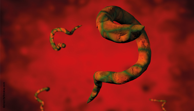
The name already sounds alarming: Echinococcoses. The diseases can be caused by infections with tapeworms of the genus Echinococcus. Basically, there are two clinical patterns: cystic echinococcosis (CE), induced by the dog tapeworm E. granulosus, which is globally spread but rarely occurs in Germany. Infection with the fox tapeworm E. multilocularis, which is prevalent at the northern hemisphere, including Germany, is the cause of alveolar echinococcosis (AE).
During their life cycles, both species change their hosts. The few millimeter small, adult tapeworms reside in the small intestine of the definitive host (carnivores, commonly dogs and foxes). Via their faeces, they release the parasite eggs which then reach the intermediate hosts through oral uptake, e.g. due to contaminated food. Intermediate hosts, required for the development of the infectious larvae may be rodents or sheep. When carnivores feed on infected intermediate hosts, the larval parasites end up in the definitive host again, where they can develop into sexually mature tapeworms. Humans are accidental intermediate hosts of both Echinococcus species. The resulting disease types, CE and AE, are characterised by the differing developmental behavior of the larvae in the human body.
Both echinococcoses are usually asymptomatic in humans for many years before they become noticeable with cholestatic icterus, abdominal pain, fatigue, weight lost and an enlarged liver (hepatomegaly). The infectious diseases may be lethal without treatment.
The larvae of E. granulosus hatch within the intestines of the infected person. Initially, they are transported to the liver via the portal vein, but may subsequently reach other organs as well. Within the tissue, the larvae form fluid-filled cysts whose slow growth is typical for the clinical course of the infection. Over the time, the growing cysts displace the surrounding tissue causing the symptoms of CE. Potential bacterial infections or rupture of the cysts and exit of fluid can lead to allergic reactions and life-threatening clinical complications.
The clinical picture of AE resembles the one of a malignant tumor. As soon as the parasite reaches the liver, the larvae develop in form of multiple, interconnected vesicles (pseudocysts). They proliferate into the tissue and destroy the organ, often unnoticed over several years. Due to the invasive growth, other organs can be attacked as well.
The diagnosis of an echinococcosis is primarily based on imaging methods which visualise the cysts. Serological tests, which detect the antibodies against the parasite in the patient sera, are performed for diagnosis confirmation. The Anti-Echinococcus-granulosus IIFT (IgA, IgM, IgG) employs frozen tissue sections of the larvae as substrate and is useful for determination of antibodies against E. granulosus, specifically. The novel Anti-Echinococcus ELISA (IgG), in contrast, is a screening test and can be used for the detection of antibodies against both E. granulosus and E. multilocularis.
