
Guest contribution:
 Article series on the subject of state-of-the-art mycology in dermatology,
13. article
by Prof Hans-Jürgen Tietz, Director of the Institute of Fungal Diseases in Berlin, Germany
I often receive questions from dermatologists describing events in their mycological mail-in laboratory. In my opinion, the establishment of local laboratory collectives is an excellent development. It’s a brilliant idea that pools strengths and is good for everyone, both colleagues and patients. Bringing together dermatological expertise in collectives for mycosis diagnostics is good for mycology and its significance. And such PCR quality circles also promote professional exchange. All this can only benefit the discipline of mycology at a time where mycoses are spreading on many fronts – whether it is the “Thailand fungus”, which has become endemic, or T. tonsurans, from tinea capitis at hairdressers to tinea corporis among wrestlers, to name just a few examples.
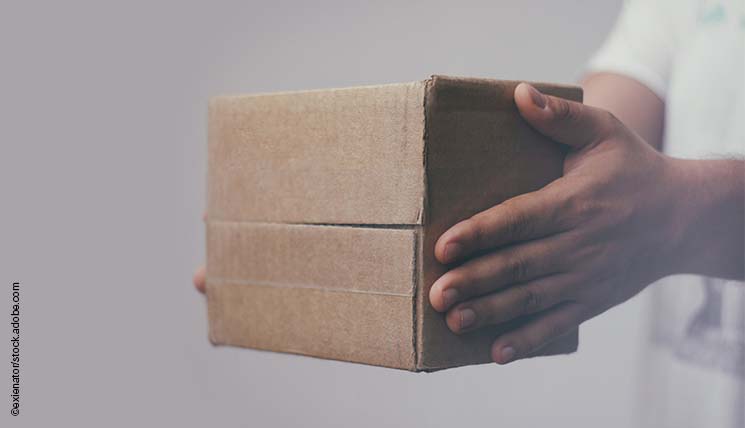
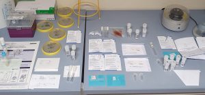
On the other hand, there has been a lack of personnel since before COVID. Medical technical assistants and receptionists who want to dedicate themselves to mycological PCRs have become rare. So the formation of laboratory collectives is also spot-on in this regard. General practitioners and paediatricians can send samples to such dermatological PCR centres, as you can see on the table in our laboratory (fig. 1). There is always quite an array of creative packaging ranging from regular envelopes to petri dishes. An ideal choice are industrial paper envelopes which can be used to deposit hair, scales or nail shavings directly from the foot or nail and are easily folded and sent by post (fig. 1 centre).
Recently, I received an email asking how it is possible that some patients have a positive PCR result for T. rubrum, although their therapy is not effective – what went wrong? In most cases, the “mistake” lies with the sender. They have to be instructed to collect a sample that is not only free of germs but also free of DNA to avoid false positive results. T. rubrum is the most common contaminant and extremely widespread. The prevalence of foot and nail mycosis is around one billion worldwide.
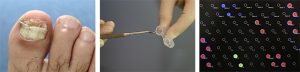
Reusable instruments such as dental hooks (fig. 2 centre) are ideal and can be freed of contaminating DNA with a thorough clean and subsequent placement in an incubator. With this procedure we have not yet had a single false positive or false negative result for our patients, which did not align with the clinical picture. But while our PCR results always aligned with the clinical picture or the therapy response of the mycosis, we also had to learn how to avoid contamination in the examination room. To collect skin scales we use commercial DNA-free swabs (fig. 1 left) or reusable sharp spoons, scissors or hooks in case of onychomycosis. Regional mail-in and consiliary PCR laboratories should regularly train their partners to avoid any misunderstanding. To aid plausibility, it is also helpful to include clinical pictures.
Another option for pathogens that are difficult to identify with conventional methods (also in quality assurance schemes) is to send them to the local dermatomycological PCR laboratory for pathogen identification. The preamble of the German Medical Association’s quality assurance regulations indicates that it must be possible for all laboratories in a quality assessment scheme to meet the requirements and that the responsible expert associations may influence the quality assurance schemes including the choice of pathogens, whose suitability must be tested “beforehand under routine circumstances using routine examination procedures” according to section E 3 1 (3).1
Be that as it may – the establishment of “PCR hotspots” in mycology are a blessing for our discipline, including in this regard. Keep it up!
Sincerely,
Hans-Jürgen Tietz
1 Dt. Ärzteblatt (2013) 110: 575-582
The articles under the rubric “Guest contribution” only reflect the views of the respective authors. Responsibility for the legality and content of the articles lies solely with the authors. EUROIMMUN does not assume liability for the completeness, accuracy and currentness of the information provided.
