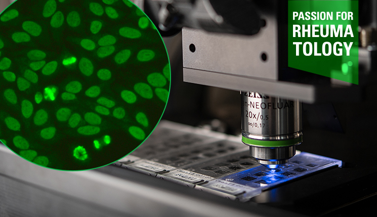
A newly published international study has demonstrated the high sensitivity of the Euroimmun Dermal Binder Mosaic 1 for detecting antibodies against laminin β4, laminin 332 and collagen type VII. The prospective multicentre study represents the latest milestone in a long-standing research collaboration between Euroimmun and the University of Lübeck, Germany.
Shedding light on anti-laminin β4 autoantibodies
Autoantibodies against laminin β4 have recently emerged as a key marker for anti-p200 pemphigoid, a form of autoimmune bullous dermatosis (AIBD) thought to be significantly underdiagnosed. In the indirect immunofluorescence assay (IFA) using salt-split skin—the standard substrate for analysing autoantibodies in AIBD—anti-laminin β4 antibodies bind to the dermal side of the artificial split. This characteristic “floor” staining pattern contrasts with the “roof” pattern produced by autoantibodies that bind to the epidermal side.
However, conventional salt-split skin IFA cannot distinguish between anti-laminin β4 and other floor-binding antibodies, such as those directed against collagen type VII or laminin 332. These are typically found in epidermolysis bullosa acquisita (EBA) and in a subset of mucous membrane pemphigoid (anti-laminin 332 MMP), respectively.
The Dermal Binder Mosaic 1: bringing clarity to the floor
The Euroimmun Dermal Binder Mosaic 1 (for research use only) enables these three floor-binding autoantibodies to be reliably differentiated. The assay consists of BIOCHIPs containing HEK293 cells expressing each target antigen—laminin β4, laminin 332, and collagen type VII—alongside control cells. By incubating all four substrates simultaneously, the test allows efficient multiparameter detection and clear differentiation of these autoantibodies.
Insights from the multicentre study
The new study evaluated the mosaic using consecutive serum samples from 41 AIBD patients showing dermal IgG binding on salt-split skin. Patients were recruited from 19 centres across 14 countries. All 41 sera reacted with at least one of the three BIOCHIP target antigens: 27 with laminin β4, two with laminin 332, and 15 with collagen type VII. No reactivity was detected in control sera from patients with pemphigus vulgaris (n=50) or healthy blood donors (n=50).
Among a control panel of 50 sera showing epidermal IgG binding on salt-split skin, three also reacted with laminin β4–expressing cells. Upon retesting, these samples revealed faint dermal staining in addition to the epidermal pattern, indicating dual reactivity. They also reacted with recombinant laminin β4 in human dermal extract by immunoblotting. These data underline the high sensitivity of the recombinant-cell IFA.
Conclusions
The authors conclude that the Dermal Binder Mosaic 1 is a valuable and useful tool for the parallel detection of autoantibodies against laminin β4, laminin 332, and collagen type VII. Moreover, the high prevalence of laminin β4 reactivity highlights anti-p200 pemphigoid as an underrecognised pemphigoid disorder.
Read the full study in the British Journal of Dermatology:
Patzelt S et al. Standardized Indirect Immunofluorescence-Based Detection of Dermal-Binding Autoantibodies in Pemphigoid Diseases: a prospective international multicentre study. Br J Dermatol :ljaf385 (2025). doi: 10.1093/bjd/ljaf385.
Learn more about the discovery and characterisation of anti-laminin β4 autoantibodies in anti-p200 pemphigoid in our previous article.