With passion for rheumatology!
Recently, three new fluorescence patterns were added to the classification tree of the International Consensus on Antinuclear Antibody Patterns (ICAP)1.
ICAP is an initiative of international scientists with the goal of evaluating the large number of ANA patterns in a standardised way. ANA (antinuclear antibodies) are biomarkers for different autoimmune diseases, such as connective tissue diseases (e.g. systemic lupus erythematosus (SLE)) or autoimmune liver diseases (e.g. primary biliary cholangitis). Therefore, ANA detection plays an important part in autoimmune diagnostics and represents the first step of serological clarification, especially in the area of connective tissue diseases. The method of choice for ANA screening is the indirect immunofluorescence assay (IFA) with human epithelial cells (HEp-2 cells). Depending on the intracellular localisation of their target antigen, ANA cause specific fluorescence patterns which may provide first hints on the specificity of the antibodies and possible disease associations.
In 2015, ICAP first published a nomenclature and classification of these patterns in order to facilitate communication between physicians, clinical laboratories and scientists.2 The patterns were classified into the main categories nuclear, cytoplasmic and mitotic. Depending on clinical relevance and differentiability, they were additionally divided into competent level (relatively easy to identify) and expert level (further differentiation). Besides their descriptive names, each pattern has been assigned a sequential code (AC, anti-cell), starting with AC-0 (negative) and AC-1 (nuclear homogeneous).
ICAP constantly updates this classification tree in cooperation with renowned scientists and clinical laboratories from all over the world. As a result of the most recent ICAP workshop, three new patterns have now been added to the existing system of 29 patterns.3
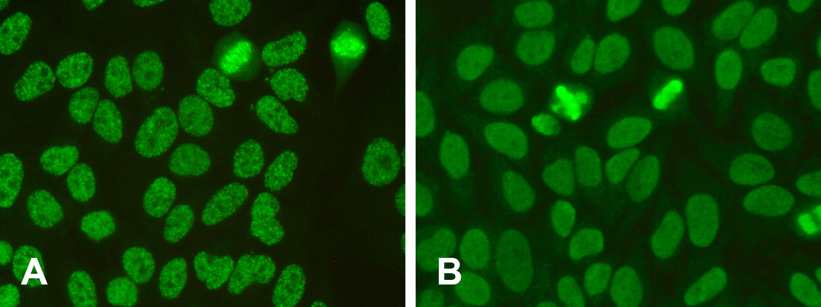
AC-30: Fine speckled with mitotic plate
AC-30 is one of these new patterns. It results from further differentiation of the pattern AC-2 “dense fine speckled”. While AC-2 is typically associated with anti-DSF70 antibodies, in several samples now classed as the new AC-30 pattern, different antibodies were detected in follow-up tests, including antibodies against nucleosomes, dsDNA, Ro/La, RNP/SM and Scl-70. Compared to AC-2, the AC-30 fluorescence pattern on HEp-2 cells presents with greater homogeneity of the speckles with respect to size and brightness, resulting in an overall “smoother” image. Both patterns share the positive reaction of the mitotic plate, which other speckled patterns, e.g. AC-4, lack. For AC-30 this staining of the metaphase is also smoother and more even. AC-30 is officially called “fine speckled with mitotic plate” and is classed as another expert-level pattern in the classification tree under the competent-level category AC-2/30 “nuclear speckled with mitotic plate”. The differentiation of AC-2 and AC-30 is also relevant from the clinical perspective: While AC-2 is associated with anti-DFS70 antibodies, which – in absence of other ANA – speak against the presence of a systemic autoimmune disease (SARD), AC-30 more often indicates presence of disease-associated autoantibodies.
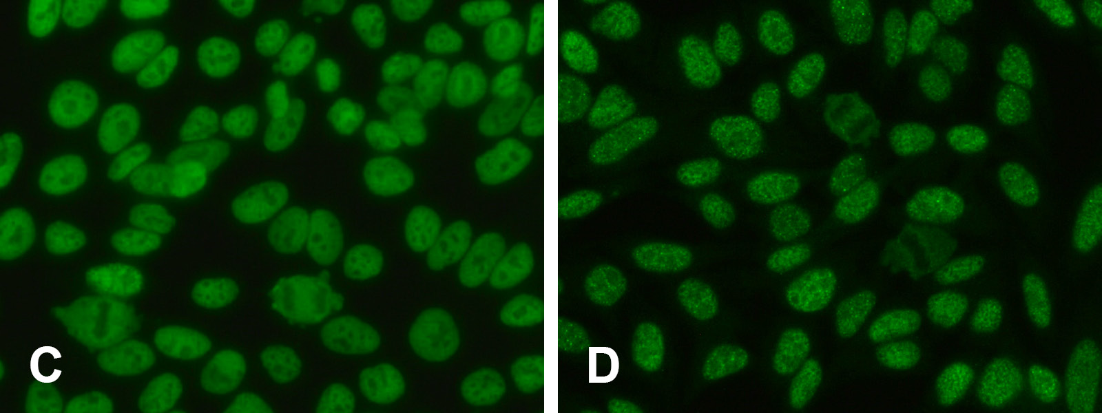
AC-31: Myriad discrete speckled
AC-31 is a variant of the AC-4 pattern that has already been described in 2013 and has now been classed as an independent new pattern. AC-31 differs from the fine and smoothly speckled nuclear AC-4 pattern by a vast number of discrete tiny dots. AC-4 occurs relatively frequently in the routine laboratory and is associated with a large range of antibodies (e.g. against SS-B/La, Mi-2, TIF1y, TIF1b and Ku). AC-31 has a strong association with anti-SS-A/Ro60-antibodies and is now classed as “myriad discrete speckled” on the expert level under the competent level category AC-4/-5/-31 “nuclear speckled”. The association of anti-SS-A antibodies with AC-31 on the one hand and of anti-SS-B antibodies with AC-4 on the other hand must still prove itself in practice, since experience shows that these antibodies often occur in parallel. Not only due to this aspect, the differentiation of AC-4, -5 and -31 (expert level) proves challenging even for experienced evaluators.
AC-XX: Unclassified
With the introduction of the pattern AC-XX, it is now also possible to record patterns that are not yet included in the ICAP-classification tree. This enables the collection of data on such patterns in order to investigate immunological and clinical links, for example in the framework of the ICAP CIC project (Clinical and Immunological Characterisation project). A more profound understanding of these patterns contributes to the advancement of autoimmune diagnostics.
The updated classification tree including all patterns is available under www.anapatterns.org and on our poster: Strategy for the determination of autoantibodies against cell nuclei (ANA) and cytoplasm components.
Despite the now 31 differentiable ANA patterns, it is usually sufficient for routine laboratory diagnostics to indicate the competent level, since subsequent confirmation and differentiation with monospecific tests is required.
2 Chan EK et al. Report of the First International Consensus on Standardized Nomenclature of Antinuclear Antibody HEp-2 Cell Patterns 2014-2015. Front Immunol. 6:412. (2015) doi: 10.3389/fimmu.2015.00412.
3 Andrade LEC et al. Reflecting on a decade of the international consensus on ANA patterns (ICAP): Accomplishments and challenges from the perspective of the 7th ICAP workshop. Autoimmun Rev. 23(9):103608. (2024) doi: 10.1016/j.autrev.2024.103608.
Click here to get to know more about our portfolio for rheumatology diagnostics. Get an overview of our range of different methods and the corresponding instruments. Together we will find the right solutions for your laboratory!